The vagus has a map. Interoception still needs a model.
posted on July 29, 2026 by Micah G. Allen
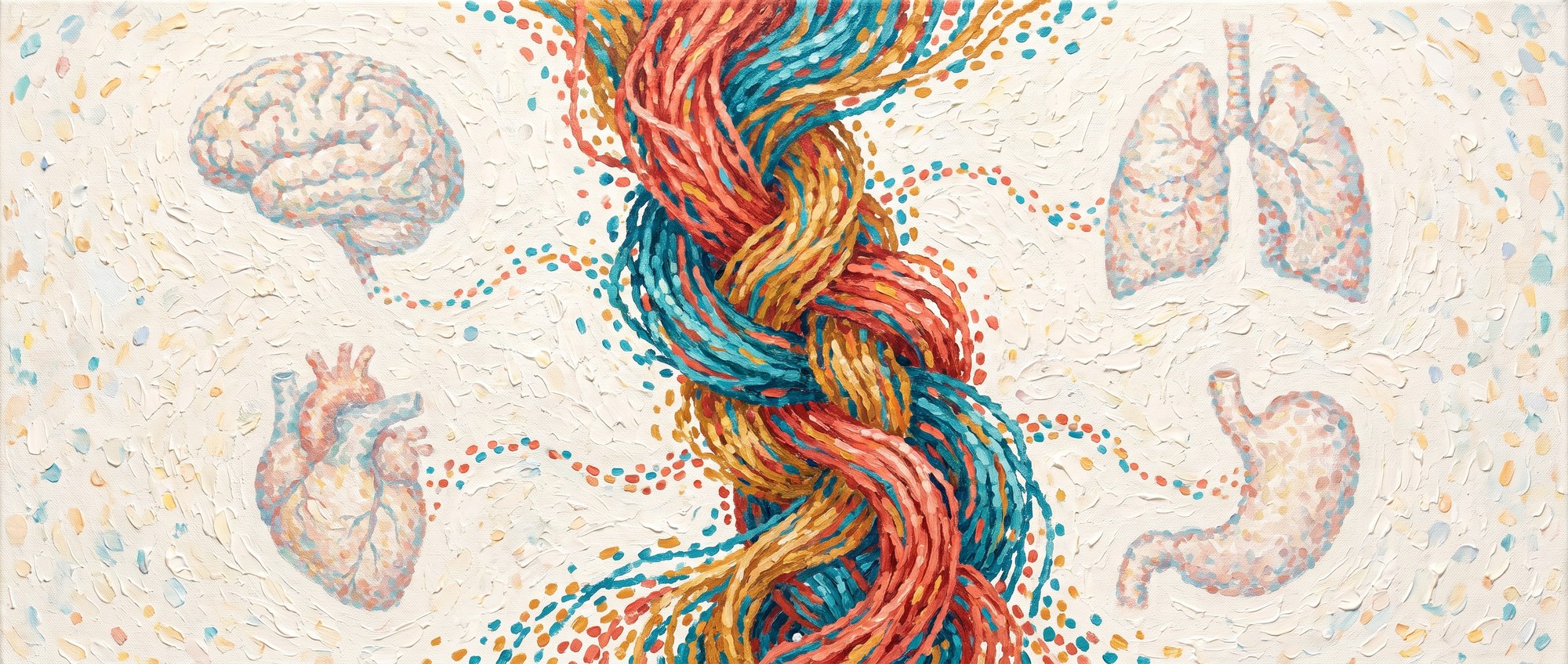
This week I was excited to see the announcement of the first population-level atlas of the human vagus nerve turn up in my feed.
The Feinstein Institutes have released a remarkable dataset of sixty vagus nerves from thirty human cadaver donors1. Using microCT, ultrasound and immunohistochemistry, they reconstructed the nerves in three dimensions along their course through the neck, thorax and abdomen. The donor-level datasets run to several terabytes each and are openly available through the NIH SPARC platform.
This gives us, for the first time, a population-level map of one of the main anatomical routes connecting brain and body. The vagus carries signals between the brain and the heart, lungs, stomach, intestines and other visceral organs, with roughly 80% of its fibres running from the body towards the brain.
Until now, most of what we knew about the organisation of individual vagal pathways came from mice. Mouse studies can identify which neurons innervate a particular organ, what those neurons respond to, and where they project in the brainstem. None of that is currently possible in living humans. The atlas arrives at an interesting moment, with mouse work becoming increasingly precise about the code just as human anatomy starts to catch up.
One question runs underneath everything that follows. Vagal signals are in some respects exquisitely organ-specific, and in others thoroughly general, and I want to know what decides which. That bears directly on how we measure interoception, how we might perturb it, and what kind of model we should be building.
A population code for interoception
The best account of vagal coding still comes from mice, and it is far richer than most people outside the field realise.
Qiancheng Zhao and colleagues barcoded the vagal sensory neurons innervating seven visceral organs in the same animal, sequenced them, and asked what those molecular identities predicted2. They found three largely independent axes. Which organ a signal came from. Which tissue layer inside that organ the nerve ending sits in. And what kind of stimulus it was, stretch or chemical or something else. Those three combine, so a modest number of genetic distinctions generates a very large number of parallel channels.
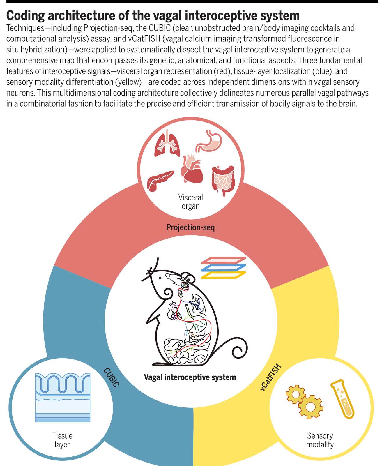
Two details have stayed with me. Organ identity turns out to be written molecularly rather than spatially, so neurons serving different organs sit intermingled in the nodose ganglion, the sensory ganglion where the vagal cell bodies live, in what the authors call a salt-and-pepper arrangement. And the projections into the brainstem run in parallel and then diverge and reconverge, with none of the tidy relay structure the textbooks draw.
This has the combinatorial depth you would expect from vision or touch. It is also the first answer to my question, and not the one I expected. A single vagal neuron is highly specific along one axis and broadly shared along another. Specificity is a property of the axis you ask about, not of the cell.
Challenges in vagal sensory mapping
The atlas speaks to one of those three axes, and only partly.
What it gives us is the human spatial substrate that mouse genetics cannot, as a distribution across sixty nerves rather than one idealised drawing. That is what you need to model what a stimulating electrode is actually recruiting in a given patient.
The limits are worth stating plainly. This is cadaver tissue from donors with a median age around 88. Press coverage has repeated a figure of 200,000 fibres in a way that suggests 200,000 axons were traced from brainstem to organ. They were not. That number is the count across both of a donor’s vagus nerves, roughly 100,000 each, and microCT at 9 µm cannot resolve a half-micron unmyelinated axon in any case. The tracing is at the fascicle level.
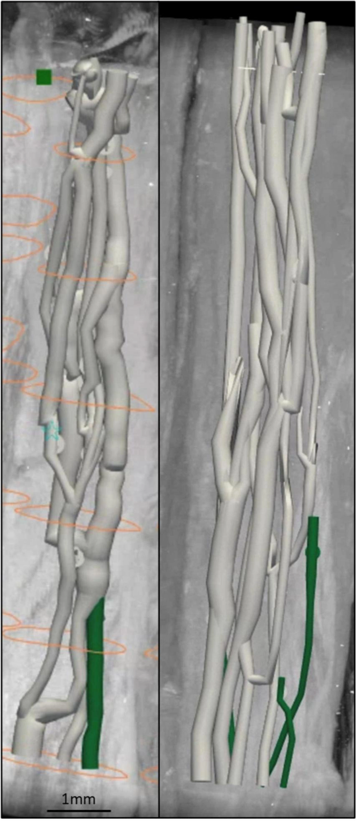
In a five-nerve pilot from David Holder’s lab at UCL, Thompson and colleagues found that cardiac fascicles are still separate from the merged trunk at the standard cuff site, which is suggestive for selective stimulation if it replicates4. At the same time the trunk reorganises roughly every 560 µm5. A fascicle you identify in one cross section has become something else a centimetre later. There is nothing stable to put a cuff around.
So anatomy can show you where fibres run. It cannot tell you which of them carry an organ-specific signal and which carry something more general, and in the cervical trunk it will not hold still long enough for you to ask.
Gastroceptive psychophysics in mice
To get at that you have to move one pathway and watch what follows. That has just become possible.
A preprint from Yoav Livneh’s group activated gut mechanosensory afferents non-invasively with optogenetics, and trained mice to report whether they had detected it6. Hits, misses, false alarms, correct rejections. Signal detection theory, on a genetically identified interoceptive pathway, in a mouse.
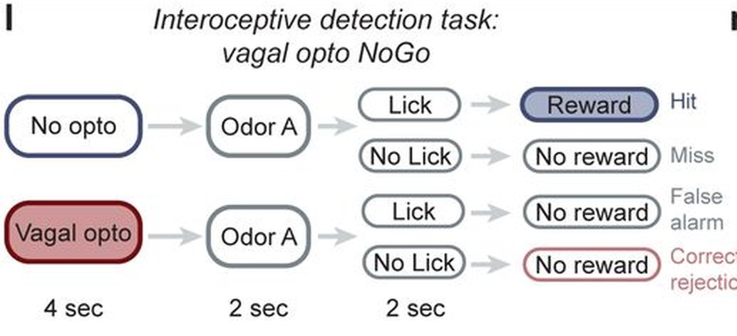
They imaged insular cortex while the animals performed the task, and the result is the one I keep returning to. Stimulus representations appeared in every version of the task. Perceptual reports appeared only in the harder psychophysical version, and manipulating insula changed behaviour only there. Whether an afferent signal becomes a perceptual report depends on what the task demands of the animal.
That is my question again, one level up. The specificity of a bodily signal is not fixed at the periphery and read out downstream. It is decided by context, and here that is shown causally. It also puts mice and humans on the same measurement footing, because detection, sensitivity and criterion mean the same formal thing in both species, so the same models can be fitted to both.
An interoceptogenic toolkit
Doing this in people needs a way to reach one pathway without opening anyone up.
Victoria Cotero, Chris Puleo and colleagues have shown, in rats and mice, that peripheral focused ultrasound can engage organ-specific autonomic pathways at chosen anatomical sites instead of at a cuff around the whole cervical trunk7,8. Ultrasound at the spleen drives the cholinergic anti-inflammatory reflex that damps cytokine release. Ultrasound at the liver moves blood glucose. A cervical cuff cannot separate those two, and with electrical stimulation the metabolic effect was originally noticed as a side effect of trying to engage the anti-inflammatory reflex. Organ-targeted ultrasound pulls them apart, and has already been through phase 1 in human spleen and liver.
Now point that beam at individual branches of the vagus. You can ask which interoceptive contributions are organ-specific and which are general, which is the experiment the coding work points at and the one nobody has run.
Focused ultrasound can also pace the heart non-invasively, at least in large animal work, which makes the cardiac signal an independent variable for once9. In human liver tumours it has already been used to release a drug inside the target organ10. Transcranially it reaches insula, which scalp-level methods do not.
The mouse toolkit is further down this road than ours. Brian Hsueh, Karl Deisseroth and colleagues built a wearable optogenetic pacemaker that sets cardiac rhythm in freely moving mice11. Induced tachycardia raised anxiety-like behaviour, but only in contexts that were already risky, and posterior insula was implicated as the mediator since inhibiting it attenuated the effect. That is cardiogenic control of an ascending signal, used to ask how it contributes to affect. Note the qualifier: only in risky contexts. Context decides again.
Put those together and you have the beginnings of an interoceptogenic toolkit, a way to write to the body-brain axis at several levels at once and without surgery. Most of it is still rodent work. It is close enough to aim at, which is why we are working to build a focused ultrasound lab in Aarhus.
Surfing the neurovisceral manifold
Tools are only as good as the model you test them against, and two results from my own lab have reshaped mine.
In matched cardiac and respiratory psychophysics across 241 people, the two axes came out largely independent in sensitivity, precision and metacognitive efficiency, with subjective confidence the one measure that tracked across domains12. In a larger sample with full symptom profiling, objective interoceptive performance was largely unrelated to mental health dimensions, while self-reported interoceptive sensibility did track symptoms13.
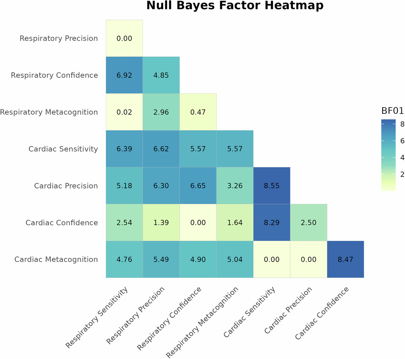
Both are positive dissociations, and each tells you something about how the system is built. In humans, cardiac and respiratory interoception are largely independent at the perceptual and the metacognitive level, so there is no single interoceptive faculty that a heartbeat task and a breathing task both index. The objective and subjective layers come apart just as strongly, with symptoms tracking what people believe about their bodies rather than what they can actually detect. If you collapse interoception into a single number, as most questionnaires do and as some of our own earlier analyses did, you throw away most of the structure worth explaining.
That structure is why I want models of joint brain-body trajectories rather than fixed channels, borrowing from manifold theory and from the dynamical systems tradition in motor control. In that framing a cardiac signal has no fixed status. At one moment a cardiac perturbation reorganises the wider network, at another it gets absorbed as one coupled fluctuation among many. So for any channel I want to know when it is driving the joint state and when it is just going along with it. There is something here about metastability that I am not the right person to develop, but the flavour seems right, and in the lab we are approaching the dynamics through integrated information and neuronal avalanches, with the aim of combining them with causal manipulations of visceral state.
A recent study in cortex seems to me to bear on exactly this question. Lorenzo Posani, Stefano Fusi and colleagues analysed more than 14,000 neurons across 43 cortical regions from the International Brain Laboratory Brainwide Map, asking whether cortical neurons sort into functionally distinct types or vary continuously14. The answer depends on scale. Across the whole cortex, selectivity is categorical and tracks anatomical connectivity. Within any single region, categorical structure is rare and largely confined to primary sensory areas, and the diversity that replaces it is what lets a downstream neuron read out many different things.
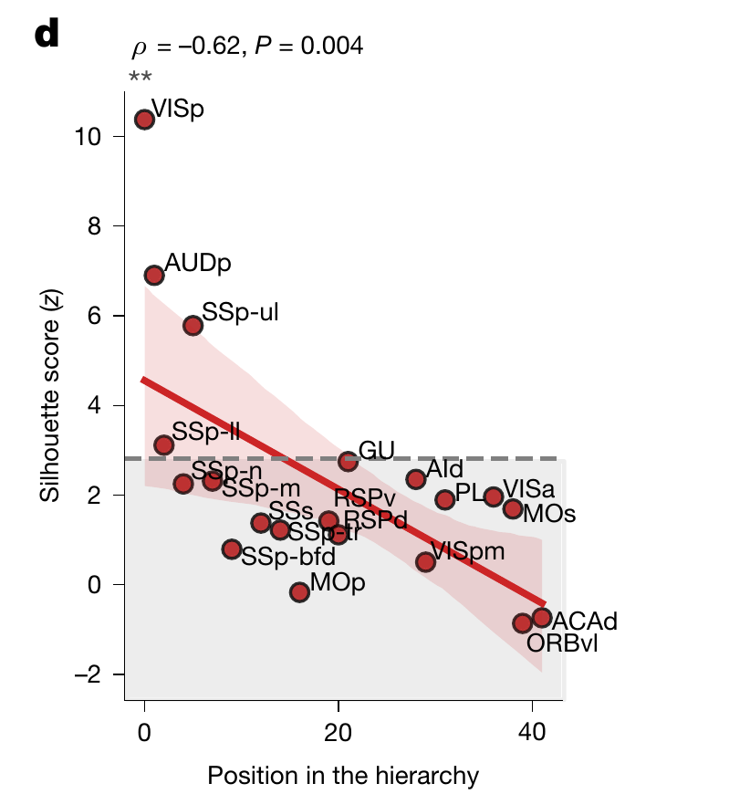
This is cortex, and mostly exteroception, so I hold it as an analogy rather than as evidence about the vagus. Still, the two results rhyme in a way I keep returning to. Zhao’s vagal neurons are specific along some dimensions and shared across others. Posani’s cortical neurons look less and less categorical the higher you go. In both cases you get a different answer about how specialised a neuron is depending on the level you interrogate it at, which is the same lesson the mouse detection task gave from a third direction.
For whom the vagus tolls
The thread through all of this is that specific and general are not fixed properties of a bodily signal. They are roles a pathway takes on, and what decides the role is context. Anatomy cannot see that. A single interoceptive score averages it away. It needs perturbation, in more than one species, fitted with models that let a channel change its status.
We can now trace the human vagus at fascicle resolution, stimulate an identified pathway in an animal that will tell us whether it noticed, pull two visceral reflexes apart with ultrasound aimed at organs, and measure several bodily axes in the same person. Five years ago I could not have written that sentence.
Most of the pieces are still provisional. But they can now be expressed in something close to a common experimental language, which means we can design experiments against the question rather than around it: when does a bodily signal govern the joint brain-body state, and when is it governed by it?
References
AI tools (Claude Code, Opus 5) were used to assist with this blog post, including literature review, figure creation, and copy editing.
Comments
Comments are threads in this site's GitHub Discussions. Posting requires a GitHub account.